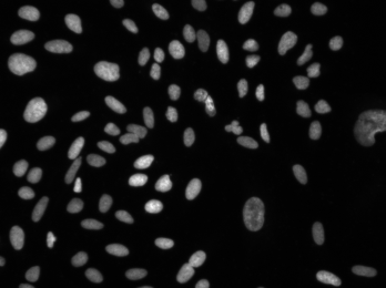
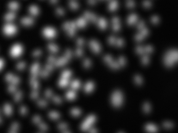
Since robust foreground/background separation and segmentation of cellular objects (i.e., identification of which pixels below to which objects) strongly depends on image quality, focus artifacts are detrimental to data quality. This image set provides examples of in- and out-of-focus HCS images which can be used for validation of focus metrics.
Images
Images were acquired from one 384-well microplate containing U2OS cells stained with Hoechst 33342 markers (to label nuclei) were imaged with an exposure of 15 and 1000 ms for Hoechst and phalloidin respectively, at 20x magnification, 2x binning, and 2 sites per well. For each site, the optimal focus was found using laser auto-focusing to find the well bottom. The automated microscope was then programmed to collect a z-stack of 32 image sets (z = 16 at the optimal focal plane, 15 images above the focal plane, 16 below) with 2 μm between slices. Each image is 696 x 520 pixels in 16-bit TIF format, LZW compression. Each image filename includes either 'w1' to denote Hoechst images or 'w2' to denote phalloidin images. *(Note: The 20x used on this particular system has an NA of 0.45.Pixel size of the camera is 6.45 um).
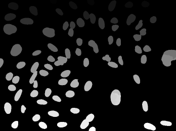
For each of the 768 fields of view (384 wells, 2 fields of view per well), an automated algorithm used the image at optimal focus (z = 16) to identify and count the nuclei (using CellProfiler’s IdentifyPrimaryObjects, applying Otsu 2-class, minimizing weighted variance; this is the likely pipeline used [download]). The counts are provided as a CSV file:
The images were manually examined by an expert to classify which focal planes corresponded to in- and out-fous images. Planes between z = 11 - 23 are considered ground truth as in-focus images.
For in-focus images, the pixels belonging to the nuclei were automatically identified as foreground and segmented (using CellProfiler’s IdentifyPrimaryObjects, applying Otsu 2-class, minimizing weighted variance; this is the likely pipeline used [download]). These ground truth images are 8-bit grayscale PNG files, with each segmented nuclei receiving a unique integer label. If the segmentation is not needed, the foreground can be thresholded with all pixel values greater than 0.
To the extent possible under law, Anne Carpenter has waived all copyright and related or neighboring rights to out-of-focus U2OS images and ground truth. This work is published from: United States.


 To the extent possible under law, Anne Carpenter has waived all copyright and related or neighboring rights to out-of-focus U2OS images and ground truth. This work is published from: United States.