Accession number BBBC010 · Version 1
Example images
 |
 |
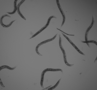
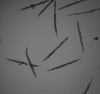
| Positive: mostly alive | Negative: mostly dead |
Biological application
This selection of images are controls selected from a screen to find novel anti-infectives using the roundworm C.elegans . The animals were exposed to the pathogen Enterococcus faecalis and either untreated or treated with ampicillin, a known antibiotic against the pathogen. The untreated (negative control) worms display predominantly the "dead" pheotype: worms appear rod-like in shape and slightly uneven in texture. The treated (ampicillin, positive control) worms display predominantly the "live" phenotype: worms appear curved in shape and smooth in texture.
For more information, please see Moy et al. (ACS Chem Biol, 2009).
Images
One image per channel (Channel 1 = brightfield; channel 2 = GFP) was acquired at MGH on a Discovery-1 automated microscope (Molecular Devices). Original image size is 696 x 520 pixels. Images are available in 16-bit TIF.
Ground truth 


For more information
These images were originally acquired for a screen in Fred Ausubel's lab at MGH. Please contact aconery AT molbio.mgh.harvard.edu for more information.
Published results using this image set
| |
|
Citation |
|---|---|---|
| 81% | 94% | Wählby et al., Nat Meth, 2012 |
Recommended citation
"We used the C.elegans infection live/dead image set version 1 provided by Fred Ausubel and available from the Broad Bioimage Benchmark Collection [Ljosa et al., Nature Methods, 2012]."
Copyright
Copyright: CC0. To the extent possible under law, the various contributors of the imagesets have waived all copyright and related or neighboring rights to BBBC010. This work is published from: United States
