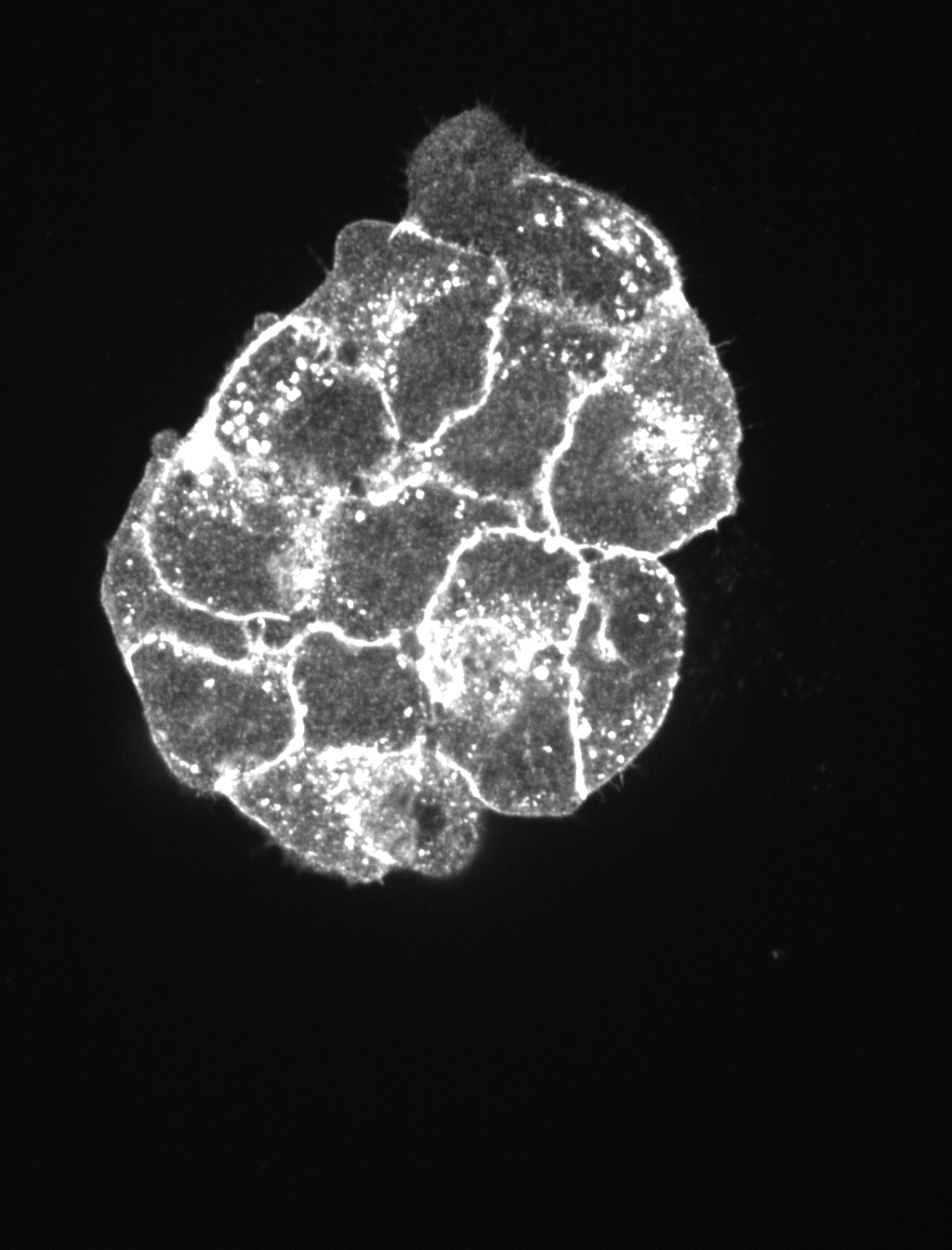
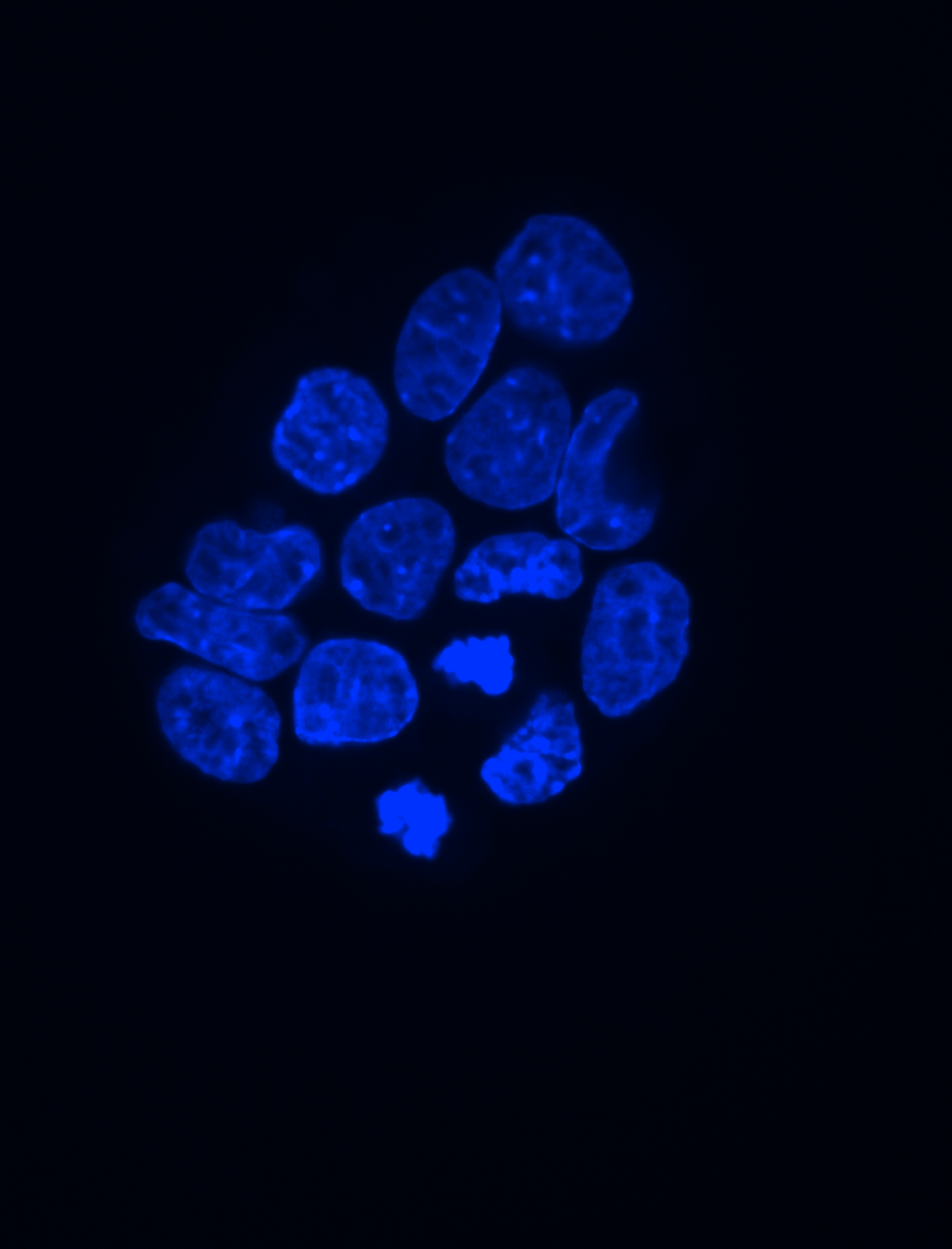
Segmenting nuclei in 3D images can be challenging especially when nuclei are clustered. Mouse trophoblast stem cells image contains a clustered monolayer of nuclei that can be difficult to segment. This image set is provided along with manually annotated ground truth that can be used for image analysis software testing purposes.
Images
Images acquired by using PerkinElmer Ultraview VoX spinning disk microscope combined with a Leica SP8 with a 63x immersion objective (Distance between Z-slices = 0.5 um)
These images were generated by the Laboratory for Modeling Development and Regeneration at Hubrecht Institute, The Netherlands. Please contact Nicolas Rivron and/or Javier Frias Aldeguer for more information.
Published results using this image set
The proposed data set will be evaluated in a publication to be submitted.


 To the extent possible under law, Nicolas Rivron has waived all copyright and related or neighboring rights to BBBC033v1.