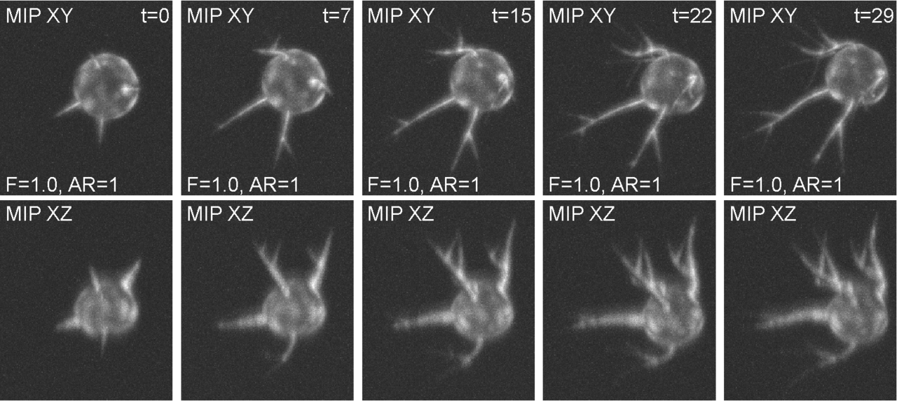
The synthetic time-lapse image data of a CRMP-2-phospho-defective cell for the given fluorescence level facto(F) and anisotropy ratio (AR).
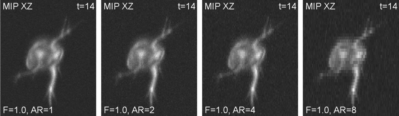
The synthetic image data of a CRMP-2-phospho-defective cell for varying anisotropy ratios (ARs).
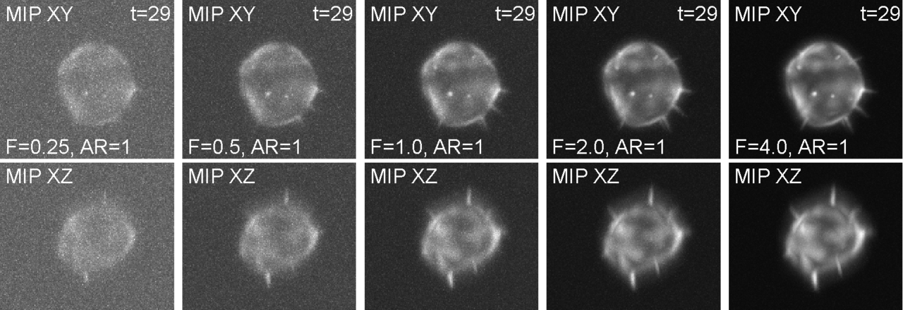
The synthetic image data of a CRMP-2-overexpressing cell for varying fluorescence level factors (Fs), which indirectly affect the signal-to-noise ratios of the image data.
Biological application
The synthetic image data displays A549 lung cancer cells of three morphologically distinct phenotypes: wild-type (WT), CRMP-2-overexpressing (OE), and CRMP-2-phospho-defective (PD). Whereas the WT cells sporadically protrude a few single-branch filopodia that may appear and disappear, the OE cells protrude many of them with short lifetimes. By contrast, the PD cells protrude aberrantly long, branching filopodia with whole-experiment lifetimes.
This image set is primarily designed to study the limits and the parameter sensitivity of fully 3D filopodium segmentation and tracking workflows with respect to the structural and temporal attributes of filopodia, such as their number, thickness, length, level of branching, and lifetime, and to the quality of image data in terms of signal-to-noise ratios and anisotropy ratios.
Images
The image set consists of 180 synthetic 3D time-lapse sequences of single A549 lung cancer cells. It includes three sequences for each of the three phenotypes, generated in 20 alternatives with five different signal-to-noise ratios and four different anisotropy ratios. Each sequence, being composed of 30 frames, was generated using FiloGen that imitates transient cell transfection with the green fluorescent protein conjugated to actin and the image formation process using a spinning disk confocal microscope equipped with a water Plan-Apochromatic 63×/1.20 objective lens.
The inherently generated reference annotations are formed of complete labeled image masks of the cell bodies and individual filopodial branches, along with comma-delimited text files describing spatiotemporal positions of filopodial tips as well as the lengths of filopodial branches over time.
M. Maška et al., "Toward Robust Fully 3D Filopodium Segmentation and Tracking in Time-Lapse Fluorescence Microscopy," 2019 IEEE International Conference on Image Processing (ICIP), Taipei, Taiwan, 2019, pp. 819-823, doi: 10.1109/ICIP.2019.8803721.
D. V. Sorokin et al., "FiloGen: A Model-Based Generator of Synthetic 3-D Time-Lapse Sequences of Single Motile Cells With Growing and Branching Filopodia," in IEEE Transactions on Medical Imaging, vol. 37, no. 12, pp. 2630-2641, Dec. 2018, doi: 10.1109/TMI.2018.2845884.