
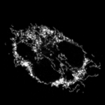
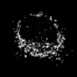
Murine Cath.a differentiated (CAD) cells-Mitochondria
Accession number BBBC053 · Version 1
Example images
|
|
Biological application
These are super-resolution images of mitochondria treated with either DMSO or CCCP to induce and detect changes in mitochondrial morphology.
Images
Mitochondria are visualized using an antibody to TOM20 and a secondary antibody conjugated to AlexaFluor 568. Images were acquired with a laser scanning confocal microscope using an Apo TIRF 60X 1.49 NA objective. Deconvolution-based super-resolution confocal microscopy was performed by acquiring z-stacks using oversampled 0.03 μm pixels and then deconvolving the images using the Landweber algorithm, resulting in images with approximately 150 nm resolution. Image size is 2048 x 2048 pixels, 16 bit .tiff format.
Ground truth 
The ground truth phenotype for each image is indicated by the directory structure.
For more information
This dataset was created by the Vitriol Lab at Augusta University, Medical College of Georgia. Please contact Eric Vitriol regarding this dataset.
Published results using this image set
Edwards, P., Skruber, K., Milićević, N., Heidings, J. B., Read, T. A., Bubenik, P., & Vitriol, E. A. (2021). TDAExplore: Quantitative analysis of fluorescence microscopy images through topology-based machine learning. Patterns, 2(11), 100367. doi.
Recommended citation
"We used image set [BBBC053] Edwards, P. et al., available from the Broad Bioimage Benchmark Collection [Ljosa et al., Nature Methods, 2012]."
Copyright
 The images and ground truth are licensed under a Creative Commons Attribution-NonCommercial-ShareAlike 3.0 Unported License (Commercial use prohibited). Eric Vitriol, evitriol@augusta.edu.