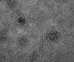
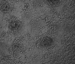
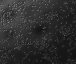
Each field of view correspond to an exposure time of LPS with 30 minute intervals between 0 hours and 30 hours. At different exposure times we expect the proportions of cell phenotypes to change, indicating the progression of the immune response to LPS.
Images
Images were collected using a Leica DMI 6000 microscope with a 20x objective, through an Andor (AND-DG-152V-C1E-FI) camera, using Metamorph Premiere Software. The images are in TIFF format.
A CSV with four columns: index, axis-0, axis-1, axis-2, and label. Each row in the CSV corresponds to a location of a cell (not necessarily centroid). 'index' indicates the annotation number. 'axis-0' indicates which FOV/image the coordinate is in. 'axis-1' indicates the x-coordinate, and 'axis-2' indicates the y-coordinate (in pixels). The column 'label' indicates the manually annotated phenotype of the cell (one of 'round', 'amoeboid', and 'stratified').
For more information
This dataset was created at Caltech, BBE, Pasadena, CA. Please contact Valentine Svensson regarding this dataset.