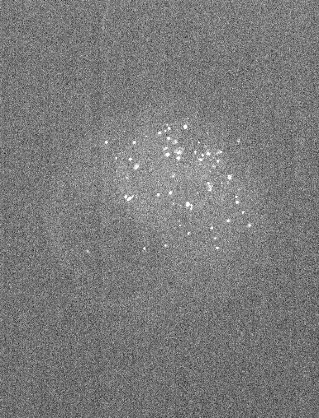
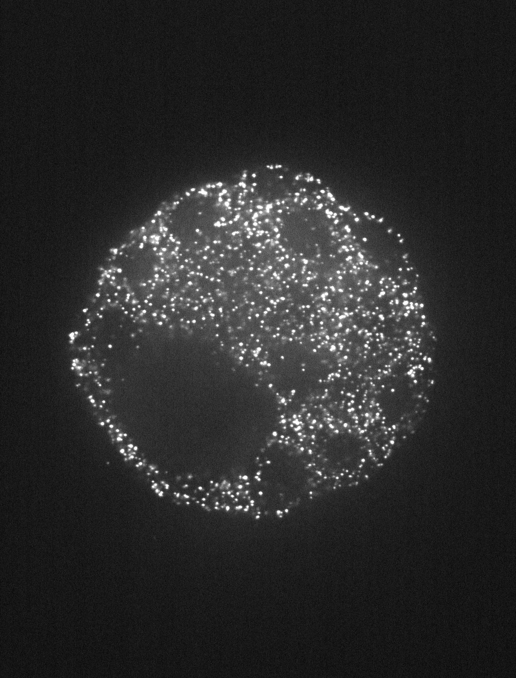
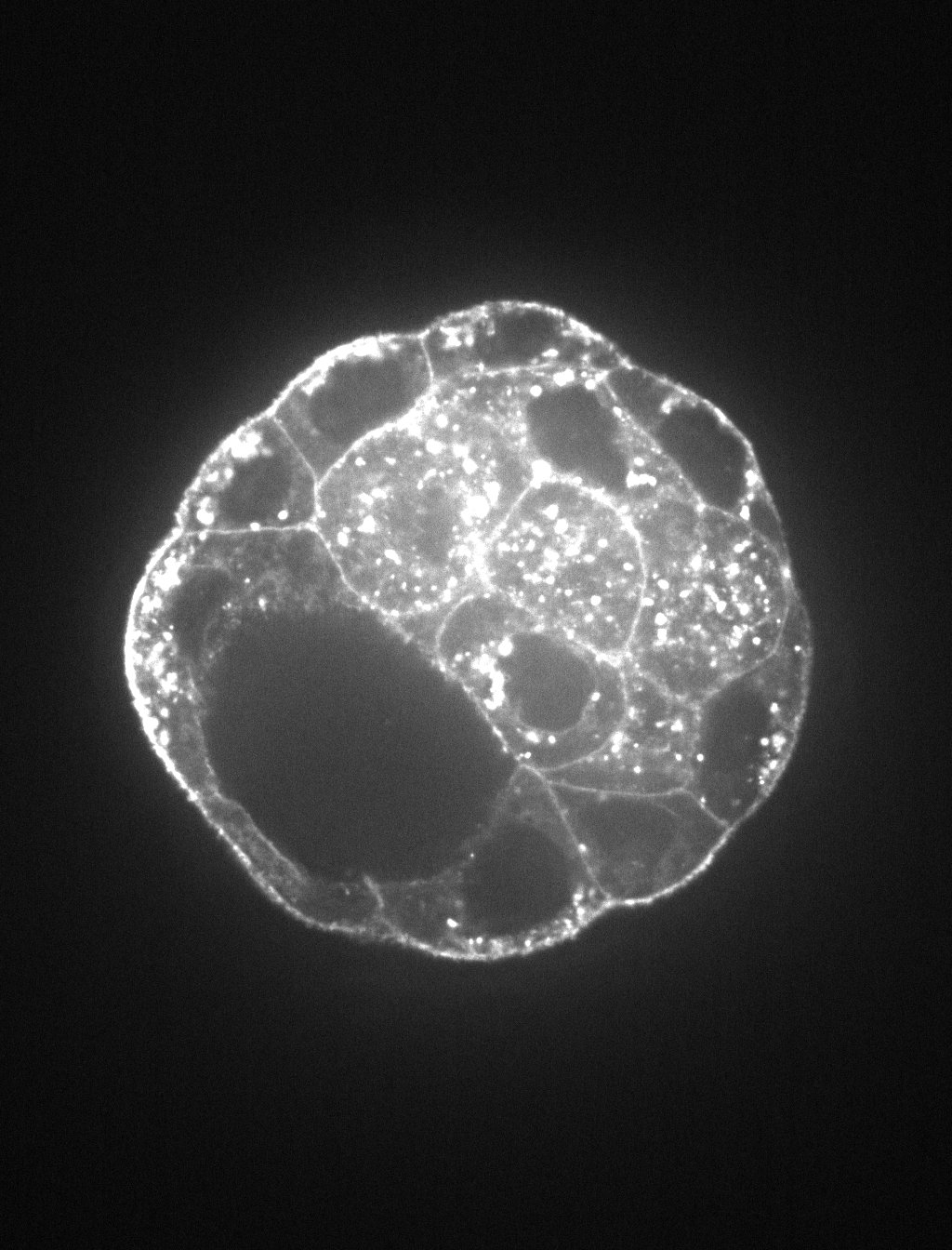
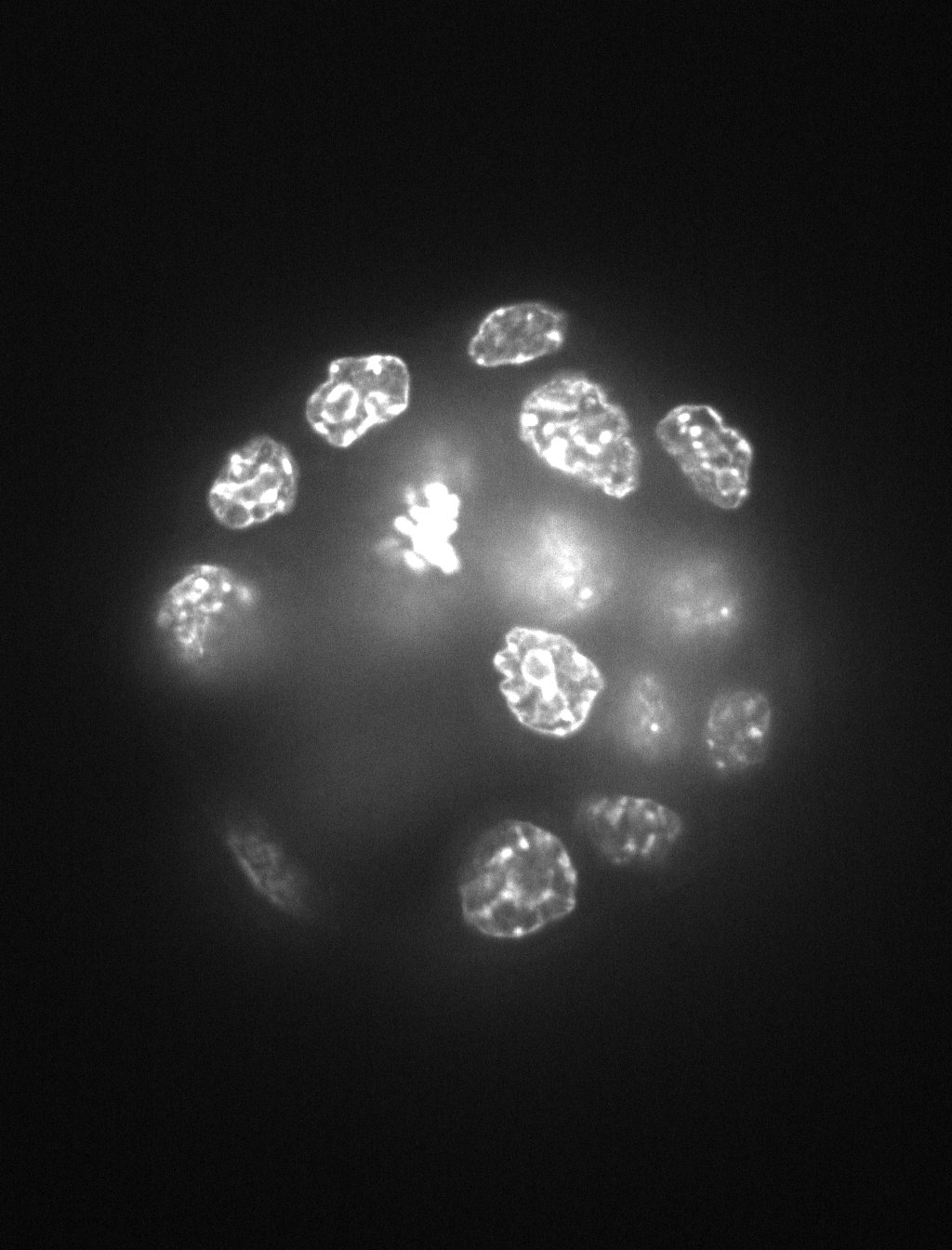
Segmenting nuclei in 3D images can be challenging especially when nuclei are clustered not only in XY plane but also in XZ and YZ planes. Manually annotated ground truth provides a reference for image analysis software testing purposes. These images of mouse embryo blastocyst cells also have changing nuclei intensity in Z plane which makes finding the right threshold for successful segmentation a difficult task. This image set also contains GAPDH transcripts that can be quantified in each cell.
Images
Images acquired by using PerkinElmer Ultraview VoX spinning disk microscope combined with a Leica SP8 with a 63x immersion objective (Distance between Z-slices = 0.5 um)
These images were generated by the Laboratory for Modeling Development and Regeneration at Hubrecht Institute, The Netherlands. Please contact Nicolas Rivron and/or Javier Frias Aldeguer for more information.
Published results using this image set
The proposed data set will be evaluated in a publication to be submitted.


 To the extent possible under law, Nicolas Rivron has waived all copyright and related or neighboring rights to BBBC032v1.